Pet Echocardiography
Images of a pet's functioning heart.
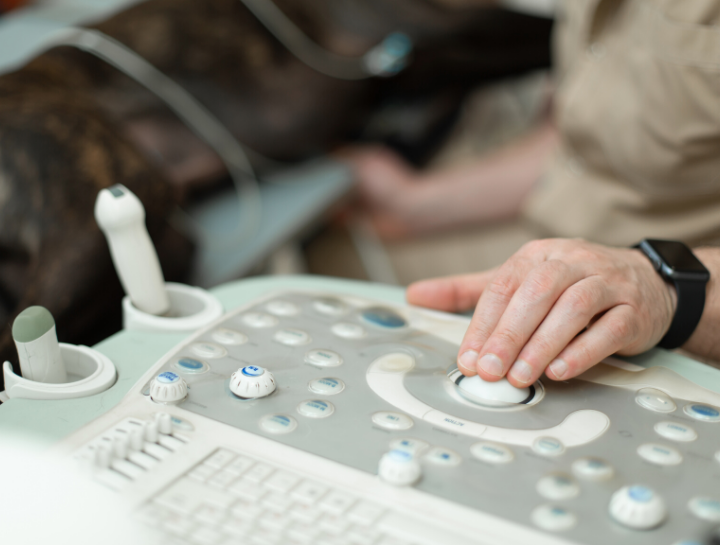
An echocardiogram is an ultrasound image of the heart and associated large blood vessels. Echocardiography, put simply, refers to an ultrasound utilized for the heart.
Echocardiography is a non-invasive test that allows a cardiologist to directly evaluate the anatomy and function of the heart. The examination is performed with the pet lying on a padded table. Most animals do not require sedation (tranquilizers) but some are more relaxed if these are used. A specific form of echocardiography called Doppler allows the cardiologist to look specifically at blood flow which helps to identify leaks in the heart valves, abnormal movement of blood between the left and right sides of the heart, and obstructions to normal blood flow.
Echocardiography is also used for diagnosis of nearly all heart diseases such as valve abnormalities (Chronic Valvular Disease), heart dilation (Dilated Cardiomyopathy), heart muscle thickening (Hypertrophic Cardiomyopathy), congenital diseases (Patent Ductus Arteriosus), heart tumors, and most other heart problems. Ultrasound for heart and abdominal scanning available once a week by a specialist with results in 24-48 hrs, special readings are available Echocardiograms, abdominal ultrasounds, pregnancy checks.
Echocardiograms can be used to detect the following conditions:
- Birth defects
- Heart disease
- Cardiac tumors
- Some heartworm infections
Like other ultrasounds, echocardiograms are painless, non-invasive, and usually don’t require any form of anesthesia. The most your furry friend will need to worry about is getting a patch of their hair shaved prior to the procedure.
If your pet has been diagnosed with heart murmurs or irregular heartbeats, call us at (609) 888-3400 to discuss whether an echocardiogram would be beneficial.